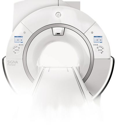
 Magnetic Resonance imaging (MRI) uses a powerful magnetic field, radio waves and computer interpretation to produce detailed images of the body’s internal structures, resulting in images that are clear, detailed and accurately characterize disease.
MRI is noninvasive and does not use ionizing radiation.
MRI can be used to evaluate the body for a variety of conditions, including tumors and diseases of the liver, heart, and bowel. It can also help doctors to see inside joints, cartilage, ligaments, muscles and tendons, which makes it helpful for detecting various sports injuries. MRI is used to examine internal body structures and diagnose a variety of disorders, such as strokes, tumors, aneurysms, spinal cord injuries, multiple sclerosis and eye or inner ear problems. It is also widely used in research to measure brain structure, and there are many other applications.
MR imaging is useful in evaluating:
- The liver, biliary tract, kidneys, spleen, bowel, pancreas, and adrenal glands
- Pelvic organs including the bladder and the reproductive organs such as the uterus and ovaries in females and the prostate gland in males
- Blood vessels (including MR Angiography)
- Physicians use MRI to help diagnose and monitor treatment for conditions such as:
- Tumors of the chest, abdomen or pelvis
- Diseases of the liver, such as cirrhosis, and abnormalities of the bile ducts and pancreas
- Inflammatory bowel disease such as Crohn’s disease and ulcerative colitis
- Malformations of the blood vessels and inflammation of the vessels (vasculitis)
Preparing for your MRI
Preparation for an MRI will vary with the specific exam. You will be given preparation instructions when your schedule your exam.
Contrast
Some MRI exams require you to receive an injection of contrast material. You must sign a consent form prior to any contrast injection. The contrast material most commonly used for MRI contains a metal called gadolinium.
Pre-Exam Screening
You will complete a screening questionnaire prior to each MRI to determine your current health status and identify if there is any material present in your body that is not compatible with MRI. Women should always inform their Radiologist or Technologist if there is any possibility that they
may be pregnant.
MRI and Metal
Jewelry and other accessories should be left at home. Because MRI uses a powerful magnetic field, electronics and any items containing metal are NOT ALLOWED in the exam room. In addition to affecting MRI images, these objects may cause you and/or others harm. These items include:
- Jewelry, watches, credit cards
- Pins, hairpins, zippers and similar metallic items
- Glasses, hearing aids and removable dental work
- Body piercings
Patients with the following implants CANNOT BE SCANNED and SHOULD NOT ENTER the MRI scanning area:
- Cochlear (ear) implant
- Some types of clips used for brain aneurysms
- Cardiac defibrillators and pacemakers
You should tell your Technologist if you have any medical or electronic devices in your body. These objects may potentially pose a risk, depending on their nature and the strength of the MRI magnet. You must provide the make and model number of any implanted device in your body.
You should notify your Radiologist or Technologist of any shrapnel, bullets, or any other metal that may be present in your body due to your occupation or prior accidents. Foreign bodies near, and especially lodged in the eyes are particularly important because they may move during the scan, and may cause blindness. Dyes used in tattoos may contain iron and could heat up during an MRI scan, but this is rare. Tooth fillings and braces are not usually affected by the magnetic field, but they may distort images of the facial area or brain, so please inform your Radiologist or Technologist of those as well.
If there is any question regarding the presence of metal or an implanted device, an x-ray will be taken to detect and identify any metal objects. In general, metal objects used in orthopedic surgery pose no risk during MRI. However, a recently placed artificial joint may require the use of MRI techniques specifically designed to image orthopedic implants.
What Will I Experience During and After My MRI?
MRI exams are typically painless. However, some patients may find it uncomfortable to remain still during imaging. Others experience claustrophobia while in the MRI scanner. If you are claustrophobic, you should speak with your referring physician about prescribing anxiety medication.
After you have been thoroughly screened for safety, you will change into a gown before being brought to the MRI room. The Technologist will position you on the moveable exam table. Bolsters and straps may be used to help you maintain the necessary position during imaging. If your physician has asked for images with contrast, an intravenous line will be inserted into a vein in your hand or arm. Coils capable of sending and receiving radio waves may be placed around or adjacent to the area of the body to be studied. Once in position, the exam table will be moved into the magnet. The Technologist will operate the MRI from a separate control room, and will be able to see, hear and speak with you at all times using a two-way intercom. You will be visible to your Technologist at all times.
You may feel slightly warm during your exam. This is normal. If it bothers you, please notify your Technologist. It is important that you remain still while the images are being obtained, which will typically be only a few minutes. When images are being recorded you will hear and feel loud tapping or thumping sounds. You will be given earplugs or headphones to dampen the sounds. You may be able to relax between imaging sequences, but will be asked to maintain the necessary position as much as possible.
MRI exams generally include multiple imaging sequences, some of which may last several minutes. If contrast material is used, it will be injected into the IV line after an initial series of scans is done without contrast.
When the exam is complete, the Technologist will check the images to determine if additional imaging is necessary. The exam table will move out of the magnet, and any coils or IV lines will be removed. Depending on the type of exam, imaging will be completed in approximately 30 minutes. You may resume normal activities and diet immediately after your exam.
Benefits and Risks of MRI
- MRI is a noninvasive and does not involve exposure to ionizing radiation.
- MRI can be more likely than other imaging methods to identify and accurately characterize diseases involving focal lesions and tumors.
- MRI is useful in diagnosing a broad range of conditions, including cancer, heart and vascular disease, and muscular and bone abnormalities.
- The contrast material used in MRI exams is less likely to produce an allergic reaction than contrast materials used for conventional X-rays and CT scanning.
- The MRI examination poses almost no risk to the average patient when appropriate safety guidelines are followed.
If you have any additional questions regarding your exam,
please call 203.337.XRAY (9729).
If you would like to schedule an appointment, click here.
MRI exams are available at the following Advanced Radiology locations
HIGH FIELD WIDE-BORE MRI:
Fairfield – 1055 Post Road
Orange – 297 Boston Post Road
Stamford – 1259 E Main Street
Trumbull – 15 Corporate Drive
Wilton – 60 Danbury Road
HIGH FIELD MRI:
Shelton – 4 Corporate Drive, Suite 182
Stratford – 2876 Main Street


